The eye is an optical instrument and vision, in part, is a physical process. Here we relate the optics of the eye to its anatomy, describe the process of accommodation and the function of reading glasses. We compare the human eye with invertebrate eyes and with a camera. This page supports the multimedia tutorial The eye and colour vision.
The eye and the camera: similarities and difference
Following parallel rays of light as they are focussed in the eye and in a camera
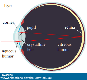
The pupil of the eye and the aperture of a camera both admit light (and both are often nearly circular). In a camera, light is focussed onto a photodetector array (or a photochemical film), typically by several lenses, which allow a zoom function and which minimise chromatic aberration. In the eye, refraction at the corneal bulge and the crystalline lens focus light onto the retina.
Anatomy and focussing
Large scale anatomy of the eye
Note that the crystalline lens is surrounded by aqueous media: the aqueous humor on the outside, the vitreous humor on the inside. These humors have refractive indices similar to that of water. This means that the crystalline lens has much less refracting power in vivo than it would have in air. Most of the refraction occurs at the cornea, the highly curve bulge on the front of the eye.
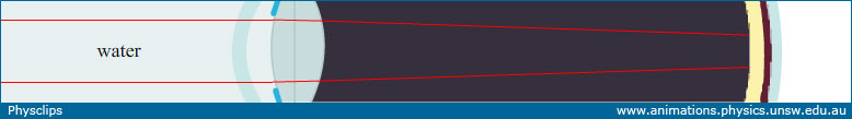
Refraction in the eye when in air (top) and in water (bottom). Without the refraction at the air-cornear interface, we are unable to focus. Swimming goggles restore this air-cornea interface and allow us to focus normally.
Camera focussing
Camera focussing
In a camera, focussing involves changing the distance between the lenses and the photodetector array. When the object is closer to the camera, the image plane is moved further from the lens. (See Lenses and images.)
Fish eye focussing
Fish eye focussing
The eyes of fish use a similar focussing mechanism: the length of the eye is increased to focus on a closer object.
Accommodation
Accommodation in the human eye: changing the shape of the lens
The human eye accommodates by changing the shape of the crystalline lens. In the Lenses and images, we saw the relation
When the object distance changes, then either the image distance changes (as in the camera or the fish eye) or the focal length changes (as in the human eye).
Loss of accommodation and the use of reading glasses
As we age, our range of accommodation typically diminishes, either due to hardening of the lens or reduction in the muscles' ability to deform it, or both. The use of converging lenses in spectacles reduces the combined focal length.
With an inflexible lens, the eye cannot focus on a close object, making reading difficult. Adding the converging lenses in reading glasses reduces the combined focal and allows focussing on the retina.
The retina and photoreceptors
The anatomy of the retina, after a drawing by Ramón y Cajal
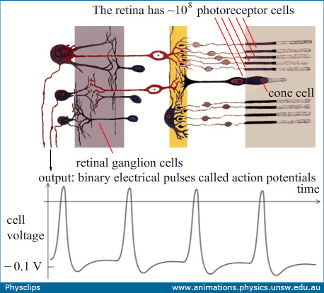
The photoreceptor cells in the human retina are classified, by their shape, into rods and cones. Cones, which are responsible for colour vision, come in three types, called red, green and blue according to whether they respond most strongly to long, medium and short wavelengths. (Page on this still to come.) Cones are concentrated in the fovea, the area directly behind the pupil, while rods are distributed over the whole retina. The photoreceptors are connected to retinal ganglion cells, which transmit electrical pulses called action potentials through the optic nerve to the brain.
In this sketch, light arrives from the left, which means that light travels through the nerves on its way to the photo receptors. This seems odd at first: in a camera, the wiring is behind the photodetector array, as shown in the photograph below. However, the 'wiring' of the eye is largely transparent.
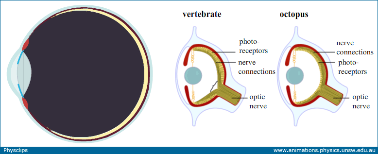
Different retinal anatomies
Comparing the vertebrate eye with that of an octopus. In the latter, the light does not pass through the nerves on the way to the photoreceptors
One consequence of having the nerves in front of the photoreceptors is the blind spot – an area in the retina without photoreceptors, where the optic nerve passes through the retina.
The blind spot
Because we have two eyes, whose blind spots are at different angles in our visual field, we usually don't notice that each eye has a blind spot. You can find your blind spot in this experiment. Close your right eye and, with the left, fixate on the X in the black rectangle (a larger version is given below). Then, move your head towards and away from the screen, always fixating on the X. When the image of the white dot falls on your blind spot, it disappears.
An experiment to find your blind spot.
The blind spot target geometry
What happens to the image on the retina?
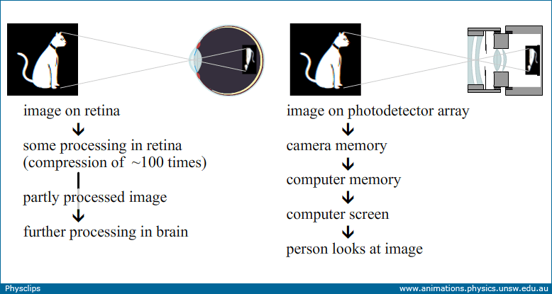
Where do the images go in the eye and the camera?
A big difference between a camera and the eye is what happens to the images. In a camera, the image on the photodetector array is usually stored, first in the camera's memory and then transferred to a computer memory, or perhaps to the memory of a photo viewer, then displayed on a screen or perhaps printed out onto paper. People look at this reproduction of the image.
Apart from optometrists and opthalmologists, no-one looks at the image on your retina, not even you. Instead, this image is processed to extract features. A small part of the processing happens in the retina itself, but most in the visual cortex of the brain. Some of this is understood: for instance, some cells respond most strongly when two adjacent regions of the retina are illuminated differently: these function as edge detectors. Others respond to more complicated patterns.